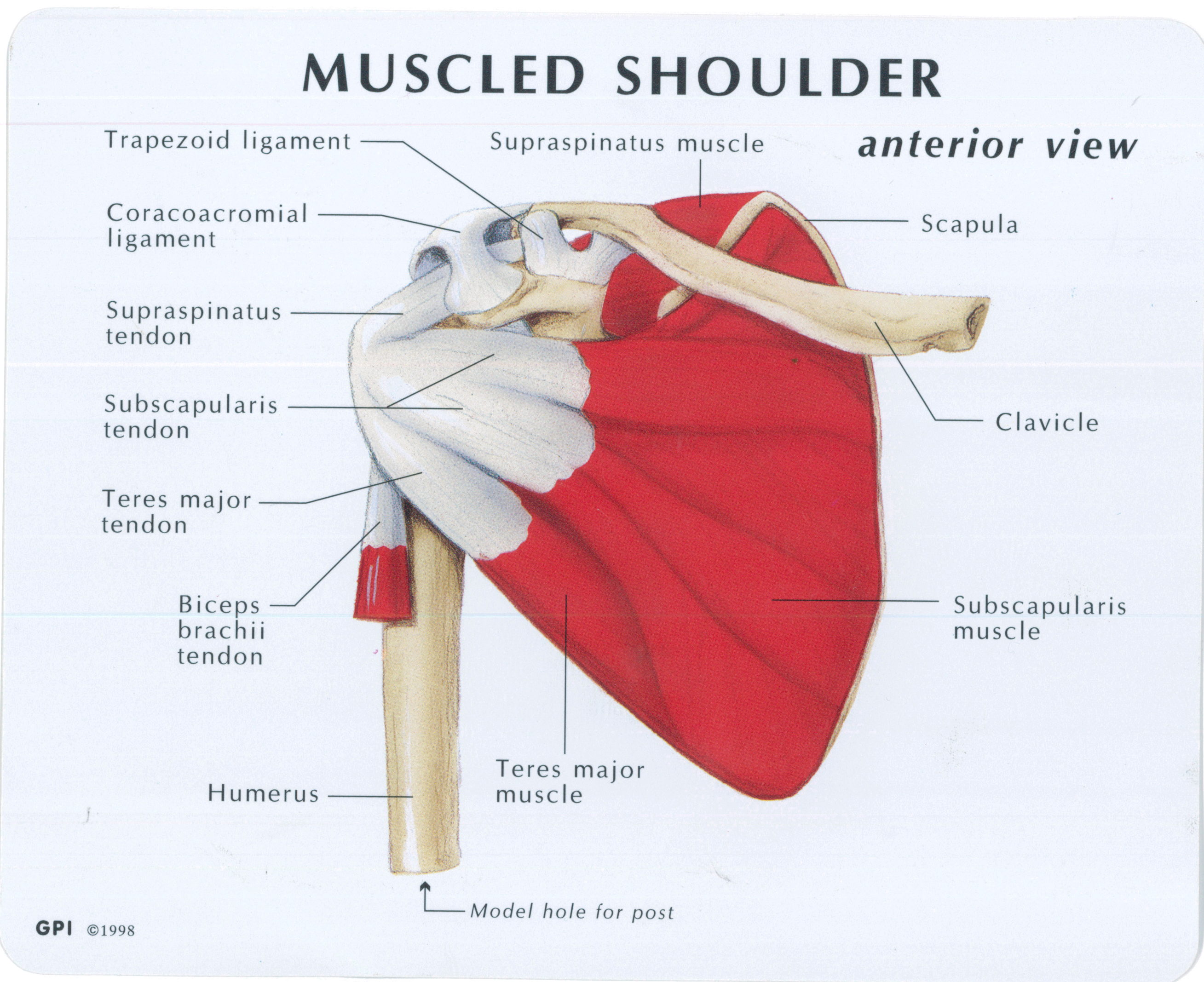
Shoulder Joint Anatomy Diagram : Diagram Of Shoulder Muscles And Ligaments - Ligaments Well Practiced Pitching Motion : Although .... Spherical end of the humerus. Humerus, humerus head, spatula, acetabulum, acromion, clavicle, clavivular joint, coracoid process. The deepest layer of the shoulder includes the bones and the joints. License image the shoulder joint ligaments shown are the acromioclavicular ligament, coracoacromial ligament, coracohumeral ligament, coracoclavicular ligament, and the articular capsule or glenohumeral. The human shoulder is the most mobile joint in the body.
Shoulder joint is the most mobile joint of the human body. It stretches across the top of the shoulder from the clavicle in the front to the scapula in the back. This incongruent bony anatomy allows for the wide range of movement available at the shoulder joint but is also the reason for the lack of joint stability. The shoulder joint by quan fu gan 60492 views. The shoulder joint is vulnerable to dislocations from sudden jerks of the arm, especially in children before strong muscles have developed.
Muscled Shoulder Joint Model - MedWest Medical Supplies from www.medwest.ca Three thickenings of the articular capsule over the anterior surface of the joint that plays very little role in strength and is mainly for joint stabilization. Atlas of the anatomy of the joint of the shoulder on a ct arthrogram in axial, coronal, and sagittal sections, on a 3d images and on conventional athrogram. 7 draw labelled diagram showing the relations of shoulder joint. Shoulder joint is the most mobile joint of the human body. See more ideas about joints anatomy, shoulder joint, shoulder joint anatomy. Erythrocyte sedimentation rate (esr) by shabab ali 20332 views. Glenohumeral joint commonly called the shoulder joint, the glenohumeral joint helps you move your shoulder forward and backward. Joint anatomy,how to draw elbow joint,elbow joint,shoulder joint,how to draw hinge joint,easy diagram,how to,how to draw ball and socket joint, how to draw hinge joint do like, subscribe, share and comment thanks for watching.
Learn vocabulary, terms and more with flashcards, games and other study tools.
The background music used in the. Describe the structure of the shoulder should begin with bone parts that include: Shoulder joint is the most mobile joint of the human body. Dislocation of the shoulder is extremely painful and may require surgical repair or even cause permanent damage. This capsule produces synovial fluid which serves to ensure. Editor · aug 6, 2017 ·. License image the shoulder joint ligaments shown are the acromioclavicular ligament, coracoacromial ligament, coracohumeral ligament, coracoclavicular ligament, and the articular capsule or glenohumeral. In this article, we shall look at the anatomy of the shoulder joint and its important clinical correlations. Three thickenings of the articular capsule over the anterior surface of the joint that plays very little role in strength and is mainly for joint stabilization. The next layer is made up of the ligaments of the joint capsule. The human shoulder is the most mobile joint in the body. The shoulder joint by quan fu gan 60492 views. Webmd's shoulder anatomy page provides an image of the parts of the shoulder and describes its function, shoulder problems, and more.
The next layer is made up of the ligaments of the joint capsule. Describe the structure of the shoulder should begin with bone parts that include: See more ideas about joints anatomy, shoulder joint, shoulder joint anatomy. The fixed joint capsule forms an envelope around the shoulder joint to seal it off from the surrounding tissue. There are several types of joints including pivot, hinge, saddle and ball and socket joints.
Anatomy of the Human Shoulder Joint from www.verywellhealth.com Erythrocyte sedimentation rate (esr) by shabab ali 20332 views. You can see it enclosing the glenohumeral joint and you can see its attachment on the anatomical neck that's the shoulder joint. Atlas of the anatomy of the joint of the shoulder on a ct arthrogram in axial, coronal, and sagittal sections, on a 3d images and on conventional athrogram. The shoulder joint is the connection between the chest and the upper extremity. Shoulder joint of human body anatomy infographic diagram with all parts including bones ligaments muscles bursa cavity capsule cartilage membrane for human shoulder joint pain anatomy. Learn vocabulary, terms and more with flashcards, games and other study tools. Just remember the articulating surfaces. Three bones come together at the shoulder joint.
The human shoulder is the most mobile joint in the body.
This capsule produces synovial fluid which serves to ensure. Equally extensive are the muscles affecting the shoulder movement, including: There are actually four joints within the shoulder: 6 describe briefly the abduction at shoulder joint. Three bones come together at the shoulder joint. The glenohumearal joint has a greater range of motion than any other joint in the body. The head of the humerus: Scapulohumeral rhythm shoulder abduction with muscular analysis. See more ideas about joints anatomy, shoulder joint, shoulder joint anatomy. This acts as the bony framework by which the muscles of the chest, upper back and shoulder connect the upper limb to the trunk of the body and control it's movements.the clavicle connects to the sternum via the sternoclavicular joint and to the scapula by. Medical and anatomical labeled scheme with. The shoulder muscles bridge the transitions from the torso. This incongruent bony anatomy allows for the wide range of movement available at the shoulder joint but is also the reason for the lack of joint stability.
There are several types of joints including pivot, hinge, saddle and ball and socket joints. This acts as the bony framework by which the muscles of the chest, upper back and shoulder connect the upper limb to the trunk of the body and control it's movements.the clavicle connects to the sternum via the sternoclavicular joint and to the scapula by. Editor · aug 6, 2017 ·. 6 describe briefly the abduction at shoulder joint. The head of the humerus:
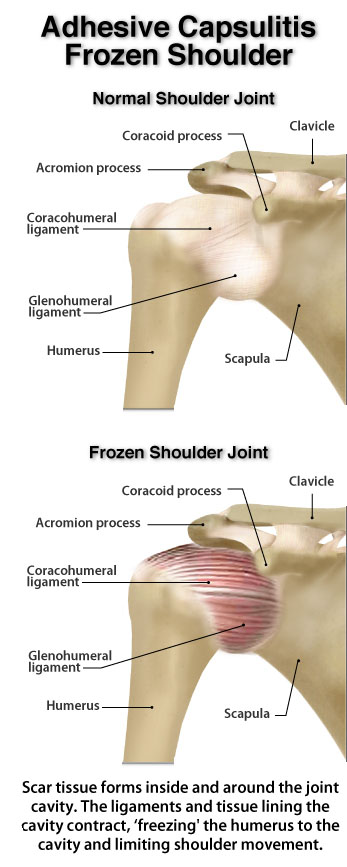
A Review of Current Frozen Shoulder Treatment Options -with Lewis Craig | POGO Physio Gold Coast from pogophysio.com.au It is the major joint connecting the upper limb to the trunk. Normal anatomy, variants and checklist. The deepest layer of the shoulder includes the bones and the joints. The fixed joint capsule forms an envelope around the shoulder joint to seal it off from the surrounding tissue. The next layer is made up of the ligaments of the joint capsule. Simple easy notes for quick revision for exams. Three bones come together at the shoulder joint. Equally extensive are the muscles affecting the shoulder movement, including:
You can see it enclosing the glenohumeral joint and you can see its attachment on the anatomical neck that's the shoulder joint.
Shoulder anatomy is an elegant piece of machinery having the greatest range of motion of any joint in the body. It has the largest range of motion out of all the joints in the body and consists of three bones. Equally extensive are the muscles affecting the shoulder movement, including: You can see it enclosing the glenohumeral joint and you can see its attachment on the anatomical neck that's the shoulder joint. Medical and anatomical labeled scheme with. Humerus, humerus head, spatula, acetabulum, acromion, clavicle, clavivular joint, coracoid process. It is the major joint connecting the upper limb to the trunk. Start studying shoulder joint anatomy. See more ideas about joints anatomy, shoulder joint, shoulder joint anatomy. 8 name the arteries and the. Learn more about the shoulder joint anatomy. Joints are the connections between bones in the human skeleton. The human shoulder is the most mobile joint in the body.
Shoulder joint is the most mobile joint of the human body shoulder anatomy diagram. The shoulder joint is the connection between the chest and the upper extremity.
Share :
Post a Comment
for "Shoulder Joint Anatomy Diagram : Diagram Of Shoulder Muscles And Ligaments - Ligaments Well Practiced Pitching Motion : Although ..."
/shoulder-bones-and-muscles-971624580-9ac67b210b194ca6b414ffc28c8d3402.jpg)

Post a Comment for "Shoulder Joint Anatomy Diagram : Diagram Of Shoulder Muscles And Ligaments - Ligaments Well Practiced Pitching Motion : Although ..."